Intraoral Camera in Johns Creek
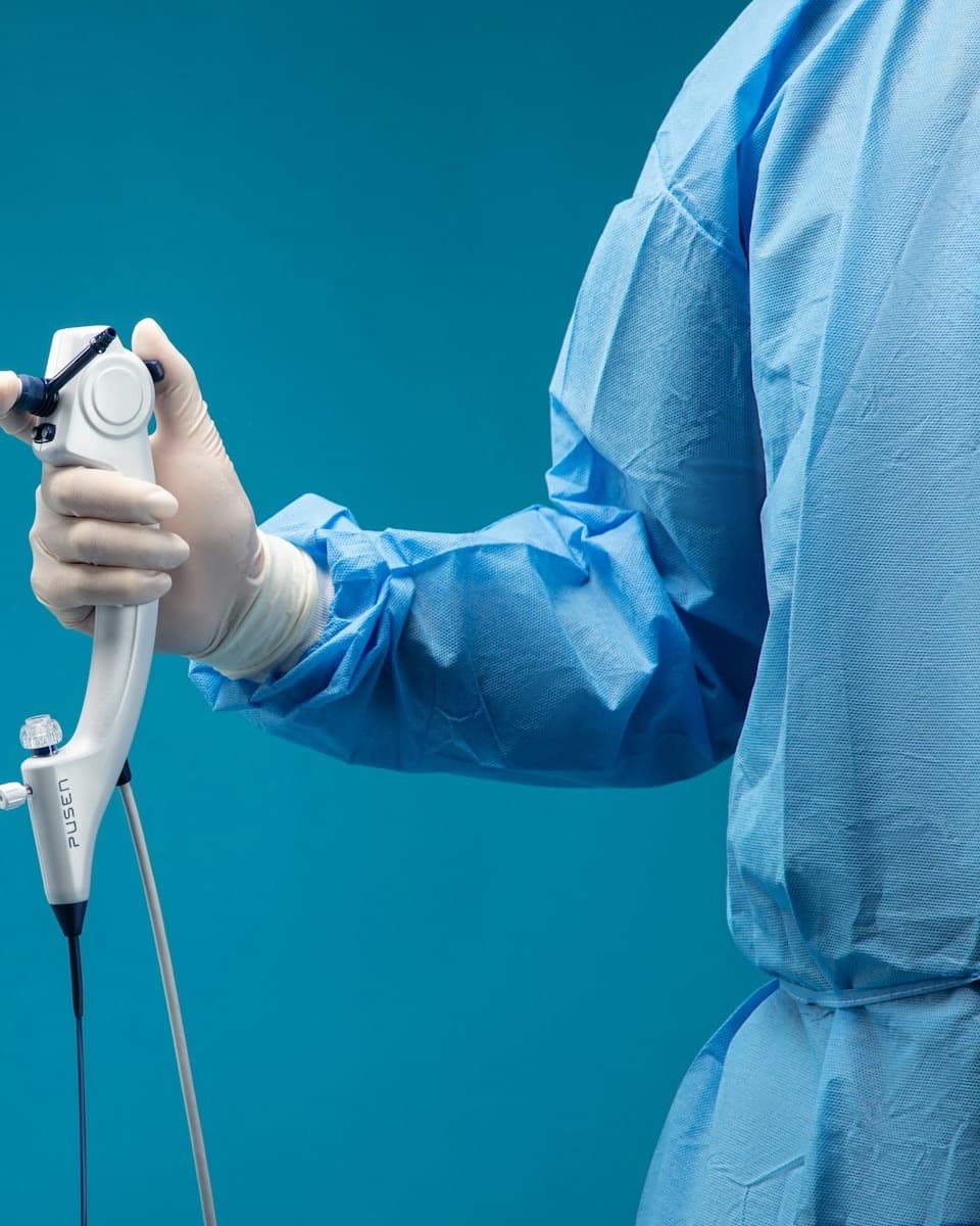
An intraoral camera is a pen-sized camera that photographs your teeth from inside your mouth and shows them on the screen in front of you. Instead of taking our word for a cracked filling or a worn edge, you see it for yourself.
- wand that fits comfortably in the mouth
- Pen-sized
- in real time, as each picture is taken
- On screen
- radiation, because it is a photograph, not an x-ray
- Zero
Curious about
the intraoral camera?
Book a consultationWhat is an
intraoral camera?
An intraoral camera is a small wand with a camera and a light at its tip. It slides into the mouth easily, and every picture it takes appears on the monitor in the room, magnified many times larger than life.
It photographs what an x-ray cannot: the surface of a tooth. Hairline cracks, worn or leaking edges on old fillings, chipped enamel, plaque you keep missing, and gum tissue that is redder than it should be all show up clearly at that size.
The pictures are saved to your chart. That means Dr. Bhoompalli can compare a tooth against how it looked at your last visit, which is how a spot that is being watched rather than filled gets tracked, and an image can be shared with you, or with your insurance company when it helps explain why treatment is needed.
At Johns Creek Family Dentistry the camera is part of how we explain things: you see the tooth on the screen, you hear what we recommend, and you decide with the picture in front of you.
How long does it take?
Seconds per tooth. Photographing a single area adds almost nothing to your visit, and even several images take only a few minutes.
Does the intraoral camera hurt?
No. The wand is about the size of a pen, rests lightly against the tooth or cheek, and there is nothing to bite on. It is one of the easiest parts of any visit.
How much does it cost?
Intraoral photos are typically taken as part of your exam rather than billed as a separate procedure. Your exact fee for any visit is set at your consultation, and we accept most insurance.
Do I need to do anything afterward?
Nothing. There is no aftercare. The images live in your chart. Ask if you would like a copy.
When we use the intraoral camera
A description is easy to doubt. A photograph is not.
The camera comes out whenever a picture will explain something better than words. It is not a replacement for x-rays, which show what is inside and beneath the tooth. The two work together: the x-ray for what is hidden, the camera for what is on the surface.
Your new patient exam
Photos of anything worth keeping an eye on, so there is a clear starting point to compare against later.
Explaining a recommended treatment
When Dr. Bhoompalli suggests a crown or a filling, you see the crack or the worn edge she is describing.
Checking old fillings and crowns
Restorations wear at the edges. Magnified, a gap or a stain line is obvious long before it becomes a cavity underneath.
A tooth you can feel but not see
Sensitivity to cold or a rough spot your tongue keeps finding often turns out to be a small chip or crack the camera can locate.
Supporting an insurance claim
When a plan asks for evidence before approving a crown or other major work, a photo can help make that case.
Tracking gum health over time
Recession and inflammation change slowly. Comparing photos from visit to visit shows whether things are improving or slipping.
Questions about the intraoral camera
What patients ask once they have seen their own teeth on the screen. Anything not covered here is a phone call away.
Still have a question? Call 770-814-7600 or text the same number.
Page updated September 2026
Is the intraoral camera the same as an x-ray?
No. An x-ray passes radiation through the tooth to show what is inside it and in the bone below. The camera is an ordinary photograph of the surface, taken with a light and a lens and no radiation at all. Each shows things the other cannot.
Is it safe for children?
Yes. There is no radiation and nothing sharp. Children tend to like seeing their own teeth on the screen, and the pictures help them understand which spots they are missing when they brush.
Can the camera find a cavity?
It can show early decay on surfaces you can see, along with cracks and stains, and it can show a filling that has started to leak at the edge. Cavities between teeth or underneath a filling still need an x-ray.
Can I get the pictures sent to me?
Ask and we will send them. The images are digital files in your chart, so we can email them to you or forward them to a specialist if you are referred.
Why photograph a tooth that feels fine?
Because the things that become expensive, a hairline crack or a filling with an open edge, do not hurt at first. A picture on file lets us watch a spot over time and treat it only if it changes.
Does the camera replace the dentist looking?
No. Dr. Bhoompalli still examines every tooth directly. The camera is for showing you what she sees at a size you can make sense of, and for keeping a record of it.
Is the camera clean?
Yes. The camera is cleaned and protected between patients, the same infection control standard we follow for every instrument in the office.
Where to next?
Related treatment
Cleanings & Exams
A cleaning every six months keeps tartar off your teeth and gum disease away, and the exam with it catches small problems while they are still small.
Explore the treatment
Related concern
Cavities or Decay
Caught early it is a filling; caught late it is more
Explore the concern
Common question
Does an early cavity need a filling or monitoring?
Learn why intact early decay and a cavity with a hole can need different care, and what to ask about monitoring or a filling.
Read the answer